2010
Mapping and data visualizations involve a process of abstraction, a distancing from the organic matter that constitutes the physical. As designers, we have at our disposal an ever increasing array of representational techniques and how, when and if we choose to adopt them unequivocally influences design outcomes and their associated aesthetics.
There is growing evidence of a trend towards the alienation of the physical and biological in our design pedagogy and an increasing interest in scales and processes which are irreconcilable with the human. It would be easy to assume that the technological precludes the experiential - indeed, it often does.
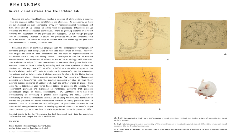
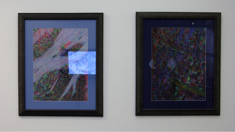
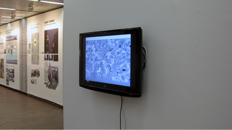
Brainbows share an aesthetic language with the contemporary “infographics” movement perhaps best exemplified in the Data Flow series of books. However, the images included in this exhibition are not maps or representations of scientific data – they are living tissue. Developed in the lab of Harvard neuroscientist and Professor of Molecular and Cellular Biology Jeff Lichtman, the Brainbow technique allows researchers to see more clearly how individual neurons connect with each other by coloring each one from a palette of about 90 shades. In this way they will be able to build up a detailed diagram of the brain's wiring, which will help to study how it computes. Unlike antecedent techniques such as Golgi stain, Brainbows operate in vivo – in the living bodies of transgenic mice. Using genetic engineering, four colors of fluorescent proteins are transferred into the genetic sequences of mice so that their neurons express mixtures of yellow, red, cyan and either orange or green. Much like how a television uses three basic colors to generate its images, these fluorescent proteins are expressed in randomized patterns that generate spectacular images of neural connections. Dr. Lichtman’s work has been revolutionary in revealing a greater (and arguably the final) layer of complexity in neural connections and his lab is using the Brainbow technique to reveal how patterns of neural connections develop in early postnatal life in mammals.
There is growing evidence of a trend towards the alienation of the physical and biological in our design pedagogy and an increasing interest in scales and processes which are irreconcilable with the human. It would be easy to assume that the technological precludes the experiential - indeed, it often does.
Brainbows share an aesthetic language with the contemporary “infographics” movement perhaps best exemplified in the Data Flow series of books. However, the images included in this exhibition are not maps or representations of scientific data – they are living tissue. Developed in the lab of Harvard neuroscientist and Professor of Molecular and Cellular Biology Jeff Lichtman, the Brainbow technique allows researchers to see more clearly how individual neurons connect with each other by coloring each one from a palette of about 90 shades. In this way they will be able to build up a detailed diagram of the brain's wiring, which will help to study how it computes. Unlike antecedent techniques such as Golgi stain, Brainbows operate in vivo – in the living bodies of transgenic mice. Using genetic engineering, four colors of fluorescent proteins are transferred into the genetic sequences of mice so that their neurons express mixtures of yellow, red, cyan and either orange or green. Much like how a television uses three basic colors to generate its images, these fluorescent proteins are expressed in randomized patterns that generate spectacular images of neural connections. Dr. Lichtman’s work has been revolutionary in revealing a greater (and arguably the final) layer of complexity in neural connections and his lab is using the Brainbow technique to reveal how patterns of neural connections develop in early postnatal life in mammals.